On Tuesday, Google announced AlphaGenome Atlas, a resource that attempts to predict the consequences of every possible single-base variant in the human genome. The human genome is about 3 billion bases long, so trying the other three DNA bases that don't appear in our reference genome means sending a total of 9 billion bases through AlphaGenome software.
AlphaGenome is designed to identify potential functions of non-coding DNA, which does not encode proteins but makes up the vast majority of the human genome. Some of this non-coding DNA is essential for controlling the activity of the protein-coding portion—it tells the cell where and when to make messenger RNAs, how to process them into mature protein-coding forms, and so on. But much of it appears to be little more than the remains of viruses and other molecular parasites.
Being able to identify the functional portion is very useful, as is having all the analysis done by a single software package. But until biologists start to use it heavily (assuming they do), it won't be clear what AlphaGenome offers beyond what we could have gotten out of its training data.
On Friday, researchers announced the completion of a map of every neuron in the brain of a male fruit fly. The "connectome" provides a tool that can accelerate neurobiology research. But it also provides an opportunity to do some science on its own, as the connectome of a female Drosophila had been completed earlier this year. The work also provided the team behind it the opportunity to refine tools that are likely to be applied to ever-more complex nervous systems, including (potentially) those of vertebrates.
The new work involved a collaboration between biologists at the Howard Hughes Medical Institute's Janelia Research Campus and computer scientists at Google—both acknowledge that neither could have done the project without the other. Preparation of an entire brain for imaging at the necessary resolution requires a distinct set of skills, as does interpreting what those images indicate. But building a complete picture of the hundreds of millions of synapses in a brain as small as the fruit fly's is a task that can't be achieved by humans in a manageable amount of time.
The people behind the effort expect that in the long term, the effort will be worth it, as the connectome could give neurobiologists a valuable tool for understanding how the brain works.
Fruit flies aren't exactly famous for their brainpower; you've probably drowned more than one in a wine glass left too long on the patio table. And yet, working with roughly 140,000 neurons—a brain smaller than a poppy seed—Drosophila can sort through a huge range of smells in a fraction of a second, and then retain the memory of that scent for a long time.
In this, they do much better than current "electronic noses." Even the most advanced ones on the market tend to be expensive, painfully narrow in what they can detect, and quick to forget an odor the moment they learn a new one.
So why not just copy the fly? That's the question a growing number of researchers have been asking—including Kevin Max and Yang Shen at the Okinawa Institute of Science and Technology, whose new algorithm, Spi-Fly, is described in a paper recently published in the journal Neuromorphic Computing and Engineering.
It's that time of year again, when we learn which lucky scientists are among the winners of the Ig Nobel Prizes. This year, the prizes honor research on designing the perfect splash-free urinal; using mosquito proboscises to "necroprint" tiny nozzles; studying composition rates of buried cotton underwear; and the aerodynamics of a healthy nose-blow, among other highlights.
Established in 1991, the Ig Nobels are a good-natured parody of the Nobel Prizes; they honor “achievements that first make people laugh and then make them think.” The unapologetically campy awards ceremony features miniature operas, scientific demos, and "24/7 lectures," whereby experts must explain their work twice: once in 24 seconds and the second in just seven words.
Acceptance speeches are limited to 60 seconds. And as the motto implies, the research being honored might seem ridiculous at first glance, but that doesn’t mean it’s devoid of scientific merit. In the weeks following the ceremony, the winners will also give free public talks, which will be posted on the Improbable Research website.
Jack Boylan, left, Allen Institute research associate, and Jesse Gray, AI BioDesign executive director of strategy and platform, at the DNA sequencer inside the initiative’s new lab. It reads millions of designed DNA sequences at once, revealing which ones worked. (GeekWire Photo / Todd Bishop)
Three of Seattle’s top scientific institutions are launching a nearly $95 million research initiative that will generate data and train AI models to design proteins and genes that don’t exist in nature — sharing the results freely to help others develop new medicines and materials.
AI BioDesign is led by David Baker, the UW biochemist who won the 2024 Nobel Prize in Chemistry for using computers to design new proteins, and Jay Shendure, a leading genome scientist at the UW and the Allen Institute.
The plan is to “hijack a lot of the machinery that evolution provided us” — the cellular assembly line that turns DNA into proteins — to design and measure millions of novel biological molecules, Shendure said in an interview in advance of the announcement.
That will help AI models learn the rules of biological design from a huge set of examples, instead of inferring them from the relatively limited number that nature has produced.
The field, Shendure said, is “putting too much emphasis on taking the cranks that we have and just running with them, as opposed to building the right cranks.”
Jay Shendure, right, lead scientific director of AI BioDesign, with research associate Jack Boylan in the lab at Dexter Yard in Seattle’s South Lake Union. (Allen Institute Photo / Jerry Petersen)
The goal is to make designing biology more like ordering a part: a molecule that latches onto a cancer cell, for example, or a genetic switch that fires only inside brain cells and nowhere else.
Potential outcomes could include everything from new therapies for disease, to proteins that dissolve plastic in the environment, to cells that travel through the body in a programmed way, said Sanjay Srivatsan, a Fred Hutch assistant professor who leads the cancer center’s work on the initiative, in a video released with the announcement.
“For the first time, the speed of AI is beginning to match the experimental power of synthetic biology,” Baker said in a statement. “That changes the question from ‘what has nature already made?’ to ‘what else is possible, and how can we test it?'”
Where the money goes
The Fund for Science and Technology is providing $94.6 million for AI BioDesign over five years. The foundation launched publicly last year with a mandate to direct a large share of Allen’s fortune into bioscience, environmental and AI research.
The funding from FFST is allocated as $46.1 million to the Allen Institute, $43.8 million to the UW and $4.7 million to Fred Hutch, according to an Allen Institute spokesperson.
The initiative had 62 people as of mid-August, including some new hires and others redirected from existing projects at the three institutions. The UW accounts for 41 of them, the Allen Institute 13, and Fred Hutch eight. AI BioDesign is expected to continue growing over time.
“AI BioDesign is exactly the kind of ambitious, collaborative science FFST was created to support,” said Marc Malandro, the foundation’s chief programs officer and co-lead, in a statement. He joined FFST in May after nearly a decade at the Chan Zuckerberg Initiative, most recently as chief operating officer of CZI and the Chan Zuckerberg Biohub Network.
Malandro and Chief Financial and Operations Officer Liz Carey have been leading FFST on an interim basis since founding CEO Lynda Stuart stepped down in May.
Inside the lab
On a recent tour of the AI BioDesign lab, research associate Jack Boylan pulled up results from a run he’d done on their new DNA sequencer that morning — on free kits donated by a neighboring biotech company, a year past their expiration date.
“We decided, let’s give it a roll,” he said. It worked fine.
The sequencer is what makes the whole approach possible. It reads all of the millions of DNA sequences in a single tube at once and reports which ones performed. One recent experiment ran 6 million distinct sequences through it at once.
“The scale comes not from robotics, but from parallelizing inside the test tube,” said Jesse Gray, executive director of strategy and platform for AI BioDesign and the Seattle Hub for Synthetic Biology, and a former Harvard Medical School geneticist.
The lab, at Dexter Yard in Seattle’s South Lake Union neighborhood, a short walk from the Allen Institute’s headquarters, is organized into teams of five or six people, each working on a different design problem.
A separate four-person team of machine-learning specialists takes the incoming results and works with the bench teams to decide which experiments come next — the ones that will teach the models the most. Each round is judged on how much the models improved.
Rui Costa, president and CEO of the Allen Institute. (Allen Institute Photo)
The Allen Institute calls projects like this “accelerators,” a term Rui Costa, the institute’s president and CEO, traced back to Paul Allen himself. The word came up in early planning sessions, Costa said. Allen wanted to “exponentially accelerate the field.”
Other accelerators at Dexter Yard include the Seattle Hub for Synthetic Biology, the Allen Institute’s collaboration with the Chan Zuckerberg Initiative and the UW, which Shendure also leads; and Cell Science, which works on engineering cells to assemble themselves into tissues.
The Allen Institute for AI (Ai2), the separate Seattle research organization also founded by Paul Allen, is involved informally rather than as a funded partner, Costa said.
Its robotics team has been talking with AI BioDesign about scaling up the protein work, and the two expect to collaborate on models and on tools that generate research hypotheses.
Why give it away
The decision to focus on open science also came from Allen, Costa said in an interview this week. “He was so visionary in the early 2000s: radically open science to exponentially impact and change fields, not to compete.”
That raises a question the initiative will face as soon as it produces anything valuable: what happens if a company builds a lucrative drug on data given away free? In traditional science, Costa said, being beaten to a discovery counts as a loss. Here it’s the goal.
“We would be so lucky if many companies would be taking this data and changing the world for good,” he said.
At the same time, Costa left open the possibility of the three principal institutions spinning out their own startups, nonprofits, or other initiatives from the work done by AI BioDesign.
Betting against the field
AI BioDesign’s approach runs against much of the current thinking in the field. Costa said most efforts to apply AI to biology are chasing a single general model that could answer questions about how any cell works. AI BioDesign is betting on the opposite: narrow models built for specific design problems, trained on data generated for that purpose.
“This project is a clear bet on a different way of doing things,” Costa said.
The people running the initiative are careful not to oversell. Gray said it remains an open question as to whether their approach beats the alternatives. “The jury’s still out,” he said.
Shendure put it plainly: “It’s never as easy as you think it’s going to be,” he said.
Costa said AI BioDesign needs to show real progress within 18 to 24 months — ideally even sooner — and expand to researchers around the world within five years.
The genetic code is what life everywhere uses to convert the information contained in DNA into specific protein sequences. With minor variations, the same genetic code is used by every living thing on Earth, suggesting it was already present in the last common ancestor of all of them. It's not an easy thing to change, because so many things in every cell depend on it.
Nevertheless, some preliminary steps have been taken. Researchers have managed to add some new amino acids to a bacterial cell and were able to make proteins that were one amino acid less than usual. But it's a slog; for some of this work, people have had to re-engineer every single gene in a bacterial genome.
Now, researchers have found a way to operate two separate genetic codes simultaneously, avoiding the need to do any work to compensate for altering the code that every protein in a cell relies on. They didn't test it in an actual cell, and it might cause some problems there. But it's a creative solution that should accelerate some synthetic biology work.
Antibodies play a central role in your body's immune defense. But they're also nearly ubiquitous in biological research, being essential for several widely used lab techniques. While some researchers need to go through the process of producing their own antibodies, antibodies to many key proteins are commercially available and can be ordered for overnight delivery.
In May, however, Reese Richardson, a post-doc at Northwestern University, discovered that some of the images used to demonstrate how these antibodies performed in lab experiments had been subject to image manipulation—the sorts of changes that would get a paper retracted if they had appeared in the academic literature. The manipulations ranged from removing background noise to copying and pasting data to fabricate results. Now, he has done a more exhaustive search and found problematic manipulations in images used to market over 17,000 commercial antibodies.
Antibodies are useful in the lab because the immune system generates them to recognize specific proteins. While those proteins typically appear on the surface of pathogens, the immune system doesn't know where a given protein it encounters comes from. So, if you inject an animal with your protein of interest, it will typically generate antibodies to recognize that.
Mummified human and animal remains have long been in demand among collectors, and such items are sufficiently rare that their acquisition may involve unregulated, occasionally clandestine trade practices—if not outright illegal ones. According to the authors of a paper published in the International Journal of Cultural Property, the risks to would-be collectors aren't just legal. Mummified remains may also pose a significant health hazard due to toxic substances used in their preservation and microbial growth due to improper storage. In fact, toxic spores or fungi are often cited as the source of the legendary "mummy's curse" tied to the excavation of King Tutankhamen's tomb.
Per the authors, the mummification process throughout history has relied on various toxic materials and substances to help preserve the remains—heavy metals like arsenic, mercury, and lead, for example—which is why careful handling is necessary to avoid adverse health effects. (Professionals typically don masks, gloves, and lab coats when working with such remains.) Various micro-organisms (bacteria, fungi, yeasts) can also lie dormant in the tissues for centuries and can recolonize and multiply due to a ready supply of organic material—particularly if the remains have been stored in suboptimal conditions.
A 1962 case, for example, revealed the presence of poisonous fungi spores in mummified remains that would cause respiratory inflammation and infection if inhaled, as well as fever. A 2013 study found that the hundreds of mummies on display at the Capuchin Catacombs in Palermo, Sicily, are prone to microbial and fungal growth after years of unstable environmental conditions. Similarly, in 2012, scientists identified three species of fungi (Aspergillus, Penicillium, and Stachybotrys) on human remains, coffins, walls, and in the air in the crypts of the Church of St. Peter and Paul in Krakow, Poland.
Our leading hypothesis for how our memories are stored is that when you learn something, the connections among neurons involved get stronger and physically larger, and that constitutes the memory. The trouble is that these connections significantly change over time—they’re plastic.
“If you compare the arrangement of these connections on day one with the same on day four or five, it's very, very different," says Kazumasa Tanaka, a neuroscientist at the Okinawa Institute of Science and Technology Graduate University in Japan. To learn how a memory that can last for years can sit on hardware that shifts every few days, Tanaka’s team made the shift a bit more dramatic. In a recent Science study, they induced a hibernation-like state in mice, which basically erased the state of more than half of their synapses. And yet the mice apparently have kept their memories.
Hibernation on demand
Hibernation is a specialty of squirrels, hamsters, and bears, but the neural circuit that triggers it is conserved across mammals, and is present in species that never hibernate in the wild—like mice. In June 2020, a team of researchers led by Takeshi Sakurai, a neuroscientist at the University of Tsukuba and a collaborator on Tanaka’s study, developed a technique to artificially activate this hibernation circuit. This can be done by activating a population called Q neurons in a region of the hypothalamus.
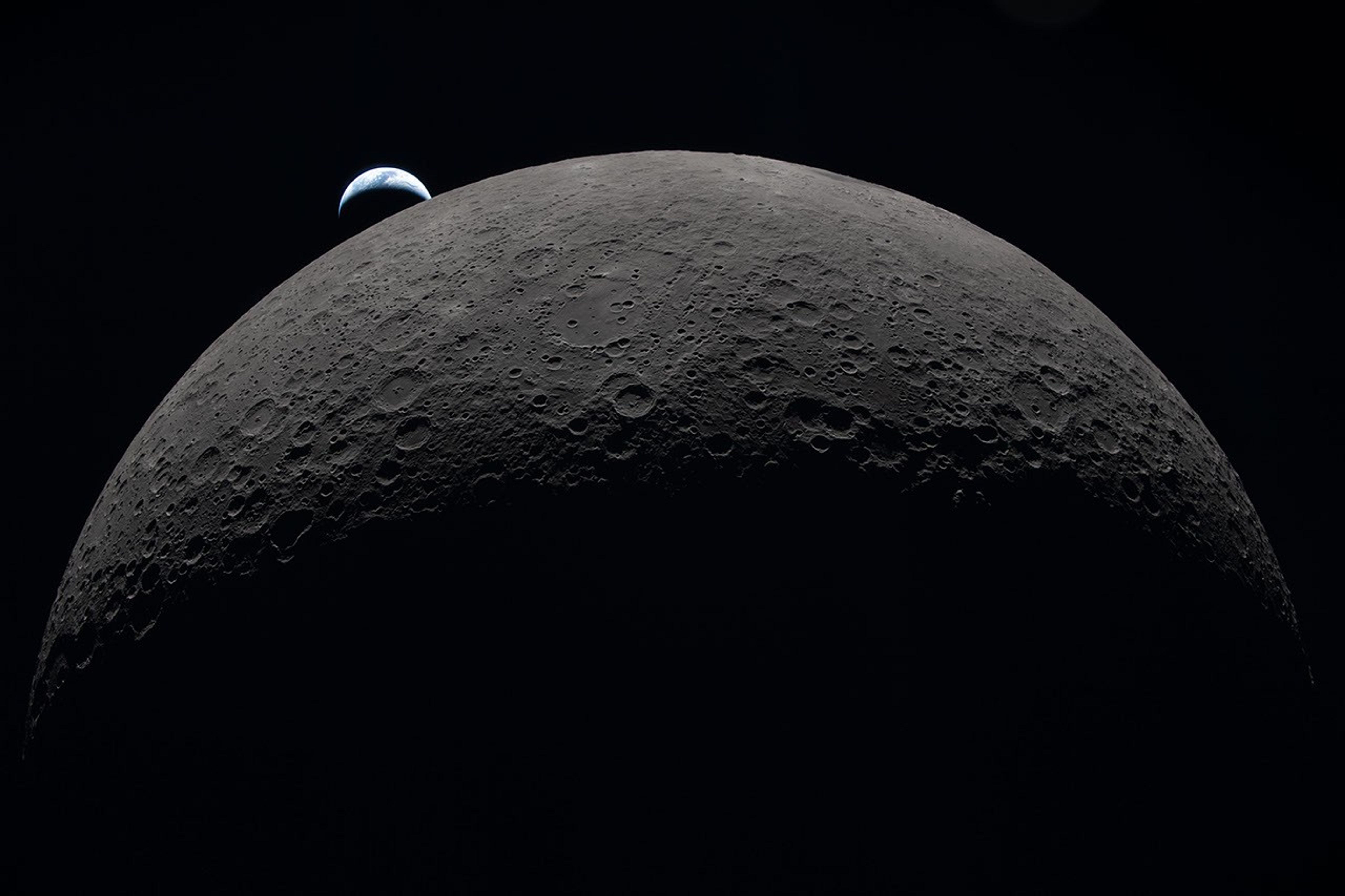
This image was taken by an Artemis II astronaut from the Orion capsule in April 2026, as the spacecraft traveled past the Moon and back over 10 days. The gray-brown, heavily cratered Moon dominates the frame against black space, with a partially lit crescent Earth setting behind its upper-left edge.
Some of Earth’s microbes likely to hitch a ride to space with human explorers could survive in the shaded nooks and crannies of the Moon’s South Pole region, NASA scientists say.
Published on Aug. 19, 2026, in Science Advances, these findings highlight a need to better understand microbial persistence in extreme lunar environments. As humans build a permanent presence on the Moon, it may become difficult to distinguish ancient lunar chemistry from contamination delivered by visiting astronauts. The concern extends beyond the Moon and on to Mars, scientists say.
“Humans are natural explorers, and with them come their voices, their memories … and their microbes,” said Prabal Saxena, a planetary scientist who led the study from NASA’s Goddard Space Flight Center in Greenbelt, Maryland. “For some scientists, myself included, that reality can be unsettling. But it also creates an opportunity to turn an imperfect situation into a useful experiment.”
Bringing microbes along is unavoidable: Humans have, on average, 1 million bacteria living on each patch of skin the size of a pencil eraser, for example. These bacteria vent from spacesuits and habitats. Though the paper’s authors worry about contamination interfering with the search for chemical clues to ancient geology or biology, they also argue that the Moon should be used as a natural lab. In shaded areas around the South Pole, scientists could carefully test the real-life limits of microbial survival in an environment that can’t easily be reproduced on Earth.
The Apollo program landed six pairs of astronauts on the Moon between 1969 and 1972. All six landing sites are near the lunar equator. In this visualization, the Apollo sites are contrasted with the South Pole, an area with enormous potential for future exploration. Time passes as we zoom toward Shackleton crater at the South Pole, revealing illumination conditions quite different from those near the equator. While many craters remain in permanent shadow, some nearby mountains and ridges are in persistent sunshine, making them attractive candidates for solar power and long-term habitation.
Before any surface science can happen, scientists need a baseline measurement of what contaminants humans bring, the authors say.
“We need to understand what was there before us, because when we go to Mars to search for signs of life beyond our planet, we will want to make sure it’s not stuff we brought,” said Andrew Needham, a NASA Goddard-based paper co-author who is an Artemis contamination‑control scientist for lunar samples.
Even with strict sterilization procedures, some organisms are stubbornly resilient. A good example is Aspergillus niger, which is a fungus that thrives in warm, damp places like household bathrooms and heating, ventilation, and air conditioning systems. Astronauts have sampled it inside the International Space Station, and experiments demonstrate that the fungus can survive outside the station as well. Aspergillus niger was one of five microbes, including bacteria and fungi, selected for this study because of its known toughness in spaceflight environments.
That microbes survived on the space station’s exterior surprised scientists. These species are typically not considered “extremophiles” that can withstand harsh conditions, such as the vacuum of space, according to Aaron Regberg, a geomicrobiologist at NASA’s Johnson Space Center in Houston.
“I would have expected these microbes to have dried out,” said Regberg, who studies space station bacteria and was a co-author on the paper.
NASA astronaut Kate Rubins on Oct. 14, 2016, collecting microbes in the Japanese Experiment Module aboard the International Space Station.
JAXA/Takuya Onishi
He pointed out that NASA often bakes robotic spacecraft at temperatures above 400 degrees Fahrenheit to reduce the number of living organisms on them. But that’s not possible with astronauts, so contamination concerns take on new meaning in crewed exploration of the Moon’s south polar environment.
A clearer picture of where microbes might survive comes from understanding how sunlight behaves at the poles. Survival in this study means the microbe can stay alive for at least one Earth day, which does not mean that it can grow and reproduce.
Because the Moon has a very small tilt on its axis, the view from its poles is of a Sun that appears to hover just above the horizon, skimming the surface like a flashlight laying on a table. As a result, elevated parts of the surface, including crater ridges, mountains, and even small bumps, block light from reaching low-lying terrain. This produces pockets of shadowed areas that can remain cold and preserve water, as well as shield fragile molecules and possible microorganisms from lethal radiation.
With that scientific context in mind, the team set out to test which Earth microbes could survive extreme polar conditions. They focused on organisms commonly found in spaceflight environments and those common on human skin. Besides Aspergillus niger, these included Bacillus subtilis, Staphylococcus aureus, Deinococcus radiodurans, and several species of Fusarium. Based on an analysis of previous studies, the scientists noted the maximum amount of heat and ultraviolet (UV) radiation each organism can withstand.
Then, the organisms were tested in simulations of three regions near the lunar South Pole — Nobile Rim, Connecting Ridge, and De Gerlache Rim. Those simulations used detailed environmental maps built from elevation and temperature data collected by instruments aboard NASA’s Lunar Reconnaissance Orbiter, combined with models of how radiation strikes the surface.
The models showed maps of “survivable niches” that range in size from a miles-wide crater floor to an astronaut’s boot print. Aspergillus niger, which was most resistant to UV radiation, was able to survive even in areas with some sunlight exposure. UV radiation is so deadly to most microbes that it’s used for sterilization in hospitals.
“When we think of the Moon, we don’t typically think of biology,” said Heather Graham, a paper co-author at NASA Goddard who helps develop tools and techniques for detecting biology that may look nothing like Earth’s. “But the Moon is a place where a cell can survive, so our first exploration of these sites should pay extra attention to our microbial hitchhikers and work hard to characterize lunar chemistry before our visits change what we will find.”
The authors note that while some microbes can survive in a dormant state in regions around the South Pole, and thereby confuse some future scientific investigations, there is no evidence the Moon has key ingredients to sustain growth and replication. Such ingredients include liquid water, which typically requires an atmosphere and moderate temperatures.
International Observe the Moon Night, Sept. 19, 2026
Each year, observers around the world come together to celebrate Earth’s Moon through direct observations, hands-on activities, lunar-themed music, artwork, readings, and more.
The Moon
From lighting up our skies to preserving evidence of our solar system’s history, Earth’s closest neighbor plays a pivotal role in the study of our planet and beyond.
About the Author
Lonnie Shekhtman
Senior Science Writer
Shekhtman helps communicate NASA planetary science to the world through news and feature stories on NASA.gov, videos for NASA+ and YouTube, and by working with the media. She reports on lunar and Mars science and exploration; NASA’s search for life; missions to Venus, Titan, and Jupiter’s Trojan asteroids; and many other topics related to NASA’s exploration of our solar system and beyond.
Scientists have long thought cynodonts, the ancestors of all modern mammals, laid eggs. For the first time, researchers have found evidence to the contrary.

 Login
Login